by Giulia Pilia1, Elizaveta Loseva1, Eliano dos Santos2, Katarzyna Kmiotek-Caller2, Ravi A. Kumar2, Anna Zoccarato2, Stuart Prime3, Jamie Bhagwan3, Svetlana Pasteuning1
1 Optics11 Life, Amsterdam, The Netherlands
2 School of Cardiovascular & Metabolic Medicine and Science, King’s College London, British Heart Foundation Centre of Excellence, United Kingdom
3 Axol Bioscience Ltd., Cambridge, United Kingdom
© 2026 Optics11 Life B.V
The first use of engineered heart tissues (EHTs) for in vitro evaluation of contractility dates back to almost three decades ago1. Since then, the development of increasingly advanced and human-derived models greatly contributed to the progress of cardiac biology2. However, the work-intensive nature of their generation, and the difficulty of retrieving accurate longitudinal measurements, has limited their use at scale. As the interest in reducing reliance on animal models for biomedical research and drug testing in favor of New Approach Methodologies grows, standardizing and increasing the accessibility to such techniques has become imperative
Hereby, we combine human iPSC-derived axoCellsTM ventricular cardiomyocytes and our high-sensitivity contractility platform Cuore into a ready-to-use solution for cardiac biology research and drug testing. Our workflow streamlines the generation, long-term culturing, and the longitudinal assessment of the contraction dynamics of EHTs. Integrating electrical pacing and preserving EHTs integrity for post-assay processing, we developed a core pipeline customizable to a broad range of applications in the cardiac biology space.
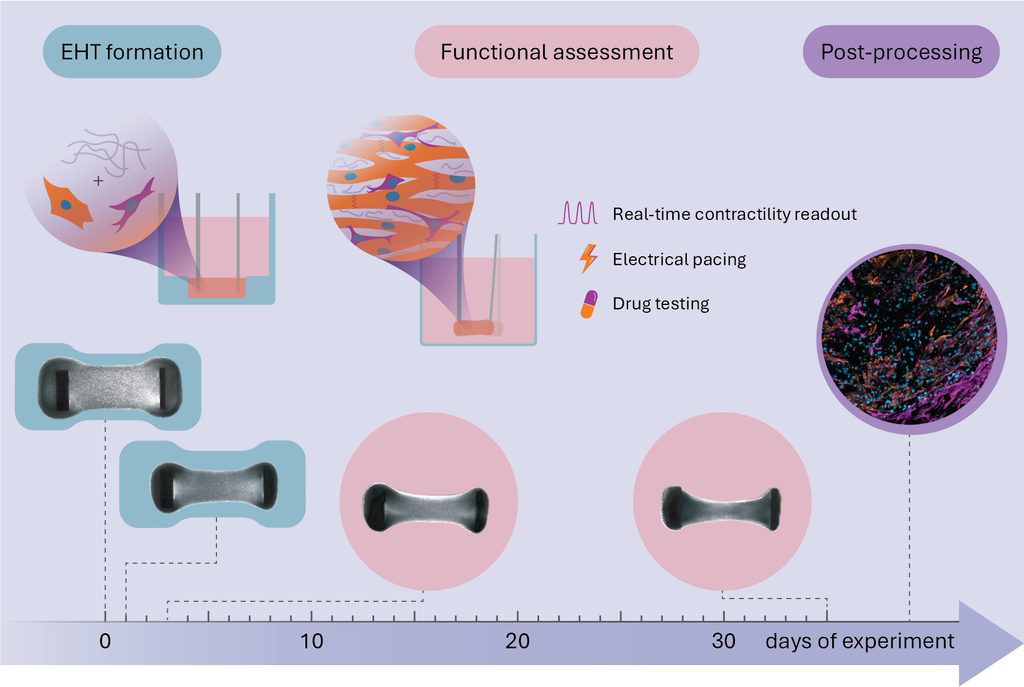
Figure 1
Overview of the presented experimental workflow. EHTs are generated in Cuore by combining collagen type I with cardiac fibroblasts and axoCellsTM cardiomyocytes. After compaction around the cantilevers, the EHTs are transferred to a standard 24-well plate. Spontaneous contractions are monitored longitudinally, and electrical pacing and drug testing can be performed. At the end of the experiment, the non-destructive nature of the measurement enables tissue retrieval for post-processing.
Results and Methods
Workflow overview
High-quality EHTs were generated following an optimized protocol inspired by Tiburcy et al.3, 4 In brief, iPSC-derived axoCellsTM ventricular cardiomyocytes were combined at a 70:30 ratio with cardiac fibroblasts (Innoprot) in a 1.34 mg/ml collagen I solution and deposited in the Cuore casting mold (Figure 1). The EHTs were cultured in Axol’s Cardiomyocyte Maintenance Medium with the addition of penicillin-streptomycin, 100 ng/ml IGF, 5 ng/ml VEGF, 10 ng/ml bFGF and – only for the first 3 days of culture – 5 ng/ml TGF-β. Medium was refreshed daily for the duration of the experiment, and the EHTs were maintained at constant gentle agitation on a rocker. On day 3, the EHTs were transferred from the casting mold to a standard 24-well plate. Spontaneous contractions were recorded daily in unstimulated conditions. When not otherwise indicated, drug effects were assessed on EHTs subjected to electrical pacing to ensure rate control.
Evolution of forces and dynamics of contraction
EHTs began to exhibit spontaneous contractions as early as day 2 from casting (Figure 2a). However, early contractions were often irregular and had elevated apparent frequency (Figure 2b). Such effect may be attributed to unsynchronous contraction across the tissue in compacting EHTs and was resolved at later timepoints. The contraction and relaxation times also varied substantially at early timepoints but stabilized by day 4 (Figure 2c). As EHTs matured, forces of contractions continued to increase, until reaching a plateau around day 15 (Figure 2a). Between day 4 and 15, while forces increased, contraction and relaxation times stayed constant (Figure 2a and c), signifying growing contraction and relaxation velocities during this period. Afterwards, forces and contraction dynamics remained consistent until the endpoint at day 36, affording a long stable experimental window.
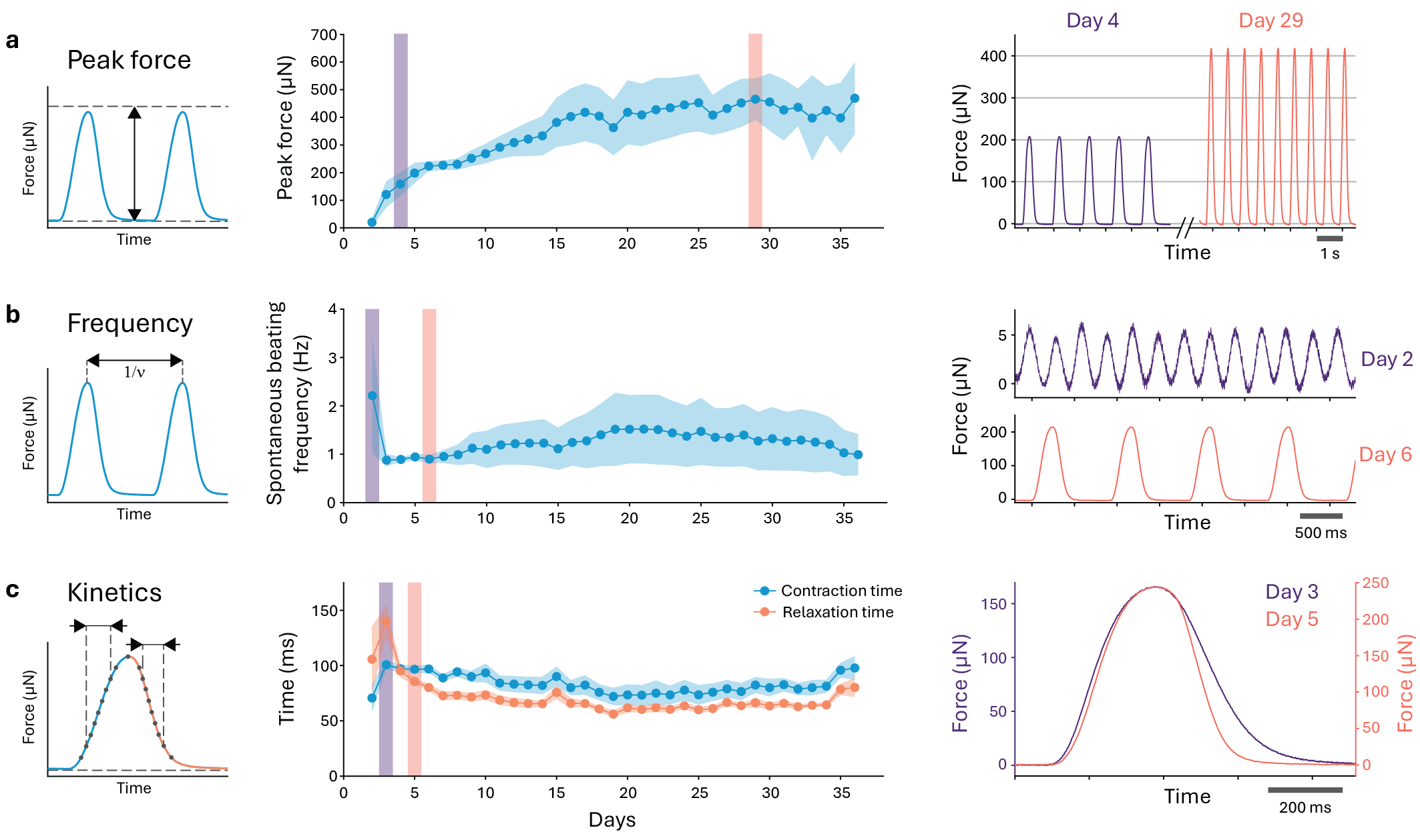
Figure 2
Evolution of spontaneous contraction parameters over the course of the experiment. (a) Peak contraction force. (b) Frequency of spontaneous contraction. (c) Time of contraction and relaxation measured as time elapsed between 20% and 80% of peak force in the ascending portion of the curve and 80% and 20% of the descending portion of the curve, respectively. In each row the right panel shows representative time-force curves for one EHT on the indicated experimental days. Solid line: mean value; shading: standard deviation (n = 12 EHTs).
Pharmacological modulation of β-adrenergic receptors
The responsivity of our model to contractility modulators was assessed by the administration of increasing doses of the β-adrenergic receptor agonist isoprenaline (Figure 3a). Contraction forces were potentiated in a dose-dependent manner, replicating the characteristic inotropic effect of isoprenaline (Figure 3b). Next, the effect of isoprenaline was modulated by co-treatment with ICI 89,406 (ICI 89) or ICI 118,551 (ICI 118), well characterized selective antagonists of β1 and β2-adrenergic receptors, respectively5. In parallel, co-treatment with a clinical stage compound (Atrogi), a first-in-class modulator of β2-adrenergic receptor hereafter named compound X, was tested (Figure 3a). Both ICI 89 and ICI 118 suppressed the inotropic effect of isoprenaline, suggesting that the EHTs in this model express both β1 and β2-adrenergic receptors.
Similarly, compound X attenuated isoprenaline’s effects (Figure 3c-d). Compound X was also effective in counteracting isoprenaline-induced effects in contraction dynamics such as reduced contraction time (Figure 3c and e) and positive chronotropic effect in unpaced conditions (Figure 3f). Notably, despite acting on the same β-adrenergic receptor subtype, compound X was more powerful than ICI 118 on reversing isoprenaline effects on both time and frequency of contraction.
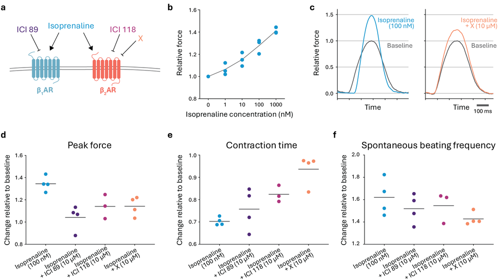
Figure 3
Effect of β-adrenergic receptors modulators on EHT contractility. (a) Schematic representation of target selectivity of the employed compounds for different β-adrenergic receptor (AR) subtypes. (b) Dose-dependent inotropic response to increasing concentrations of isoprenaline. (c) Representative force-normalized contraction curves of EHT before and after treatment with the indicated compounds. (d) Peak force. (e) Time of contraction between 20% and 80% of peak force. (f) Frequency of spontaneous contraction. Dots: single EHT value; line: mean value. The contractility parameters are normalized, for each EHT, to the value before drug administration.
Conclusions
- Cuore’s measurement capabilities, combined with axoCellsTM ventricular cardiomyocytes provide a complete solution for the modelling and longitudinal monitoring of human cardiac function in vitro.
- In our model, tissue contractility parameters such as force, frequency, and kinetics of spontaneous contraction evolve towards a stable phenotype, affording a broad experimental window for longitudinal studies.
- As exemplified by modulation of β-adrenergic receptors with both reference and test compounds, the presented setup enables drug testing both in unstimulated and rate-controlled conditions, establishing a complete versatile platform for cardiac biology research and therapy development.
Acknowledgements
We acknowledge Atrogi AB (Sweden) for providing compound X and for sharing expertise in adrenergic receptor biology.
References
- T. Eschenhagen et al. Three-dimensional reconstitution of embryonic cardiomyocytes in a collagen matrix: a new heart muscle model system. FASEB J. Off. Publ. Fed. Am. Soc. Exp. Biol. 11, 8, 683–694 (1997).
- I. Gokhan, T. S. Blum, and S. G. Campbell. Engineered heart tissue: Design considerations and the state of the art. Biophys. Rev. 5, 2, 021308 (2024).
- M. Tiburcy et al. Defined Engineered Human Myocardium With Advanced Maturation for Applications in Heart Failure Modelingand Repair. Circulation. 135, 19, 1832–1847 (2017).
- M. Tiburcy, T. Meyer, N. Y. Liaw, and W.-H. Zimmermann. Generation of Engineered Human Myocardium in a Multi-well Format. STAR Protoc. 1, 1, 100032 (2020).
- J. G. Baker. The selectivity of β-adrenoceptor antagonists at the human β1, β2 and β3 adrenoceptors. Br. J. Pharmacol. 144, 3, 317–322 (2005).