Spinal cord tissue is mechanically complex, with properties that depend on tissue type, testing conditions, spatial scale, and loading rate. Understanding this complexity is important for predictive medicine, neurosurgical planning, disease modeling, drug delivery simulations, and regenerative strategies.
In this Faces of Mechanobiology entry, Oskar Neumann from Friedrich-Alexander-Universität Erlangen-Nürnberg explores how to measure spinal cord mechanics across scales. He explains why reported stiffness values in the literature can vary widely, how Chiaro enables tissue-specific characterization of gray and white matter, and why combining mesoscale indentation with macroscale mechanical testing can support more robust material parameters for computational models of spinal cord biomechanics.
Why spinal cord mechanics needs scale-aware measurement
The mechanical signature of central nervous system (CNS) tissue, particularly the brain and spinal cord, is increasingly recognized as relevant to predictive medicine, clinical applications, and therapeutic strategy development. Potential applications range from drug-delivery planning and disease-progression simulations to neurosurgical planning and intraoperative guidance. A better understanding of the time- and scale-dependent mechanical properties of CNS tissue is therefore an important driver of innovation across these fields.
Within the Collaborative Research Center “Exploring Brain Mechanics,” Project B01 focuses specifically on spinal cord mechanics and the role of mechanical cues during regeneration, which has recently been shown to be closely linked to changes in tissue mechanics in regenerating species. To understand the interplay between mechanics, biology, and biochemistry during spinal cord regeneration, a thorough characterization of the underlying mechanical properties of spinal cord tissue is essential. However, the existing biomechanical literature reports highly inconsistent stiffness values, largely due to differences in testing techniques and experimental conditions, including post-mortem time, sample preparation, and testing protocols. Even among indentation-based studies, reported stiffness values can differ by several orders of magnitude.
Mesoscale indentation of gray and white matter
Coming from the field of continuum mechanics, our group employs several experimental approaches to quantify the mechanical properties of spinal cord tissue. A key step toward understanding the mechanical differences between gray and white matter is the use of mesoscale indentation, which enables tissue-specific characterization at an appropriate spatial scale. Using the Chiaro Nanoindenter, we tested porcine spinal cord gray and white matter (Figure 1) and observed the rate-dependent viscoelastic behavior characteristic of CNS tissue. We further found that environmental and experimental conditions, including temperature, test duration, and particularly the fluid in which samples are submerged during indentation, can substantially influence the measured mechanical properties [1].
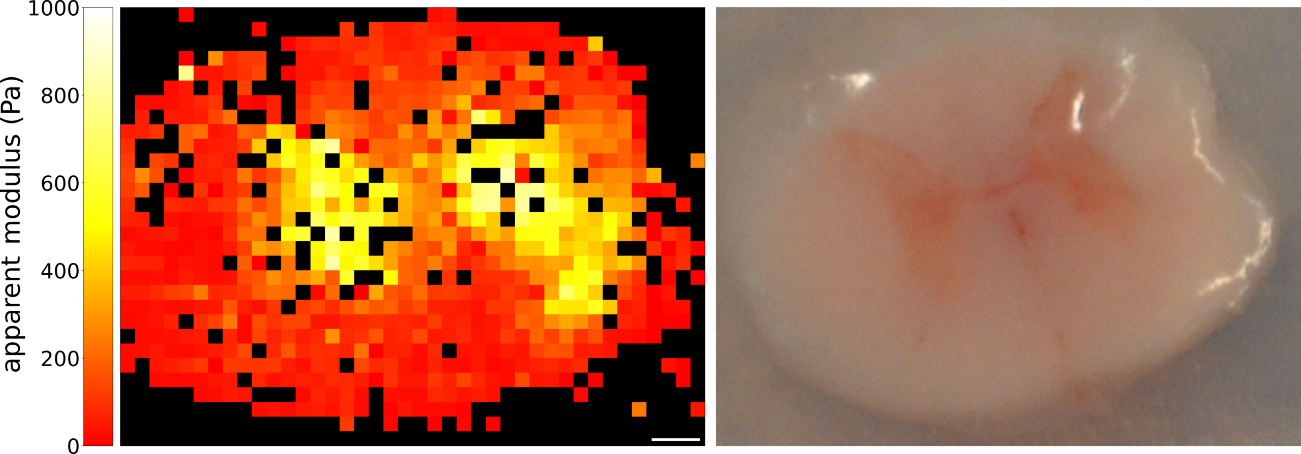
Figure 1. Heat map of the apparent modulus of one exemplary spinal cord transverse plane, with stiffer areas highlighting the characteristic shape of spinal cord gray matter tissue on the left side. The scale bar denotes 1 mm. Image of the corresponding spinal cord slice on the right side.
How indentation scale changes what we see
Further, to elucidate contradictory findings in the literature, we developed a multi-bead testing protocol in which the exact same locations in gray and white matter were tested subsequently using nominal bead diameters of 20, 100, 200, and 500 μm [2]. In agreement with the general trend observed across the literature, averaging across technical and biological replicates revealed a clear shift in the gray-to-white matter stiffness ratio (Figure 2a). At smaller indentation scales, gray matter exhibited higher apparent moduli, whereas larger indenter beads resulted in higher apparent moduli for white matter. This turnover was observed in the transverse plane, while no comparable tendency emerged in the coronal plane. These findings highlight the complex mechanical nature of spinal cord tissue, characterized by both anisotropy and scale dependence. We hypothesize that this scale-dependent response reflects the anisotropic axon–glia architecture of white matter, with larger indenters engaging a broader mechanically coupled network, as illustrated in Figure 2b and c.
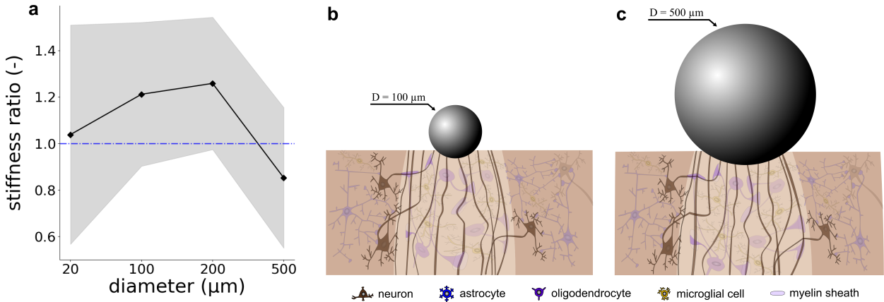
Figure 2. (a) Ratio of spinal cord gray to white matter stiffness shifts between indenter diameters of 200 and 500 μm across Protocols. Mean ratio values of all biological replicates across different indenter diameters and protocols in black, with standard deviation in gray. The blue dashed line indicates a ratio of one. (b-c) Schematic of deformation state during spherical indentation. Spinal cord white matter tissue is shown with axonal tracts, myelinating oligodendrocytes, and microglia; myelin sheaths are depicted only exemplarily on selected axons. Spinal cord gray matter is shown with neurons, protoplasmic astrocytes and microglia cells. Both schematics are illustrative and not to scale.
Toward multimodal spinal cord characterization
To integrate our indentation-based mechanical characterization of spinal cord tissue into a more holistic parameter identification framework, we combined tissue-type-specific indentation with whole-sample rheometer-based compression–tension testing in a multimodal experimental pipeline (Figure 3).
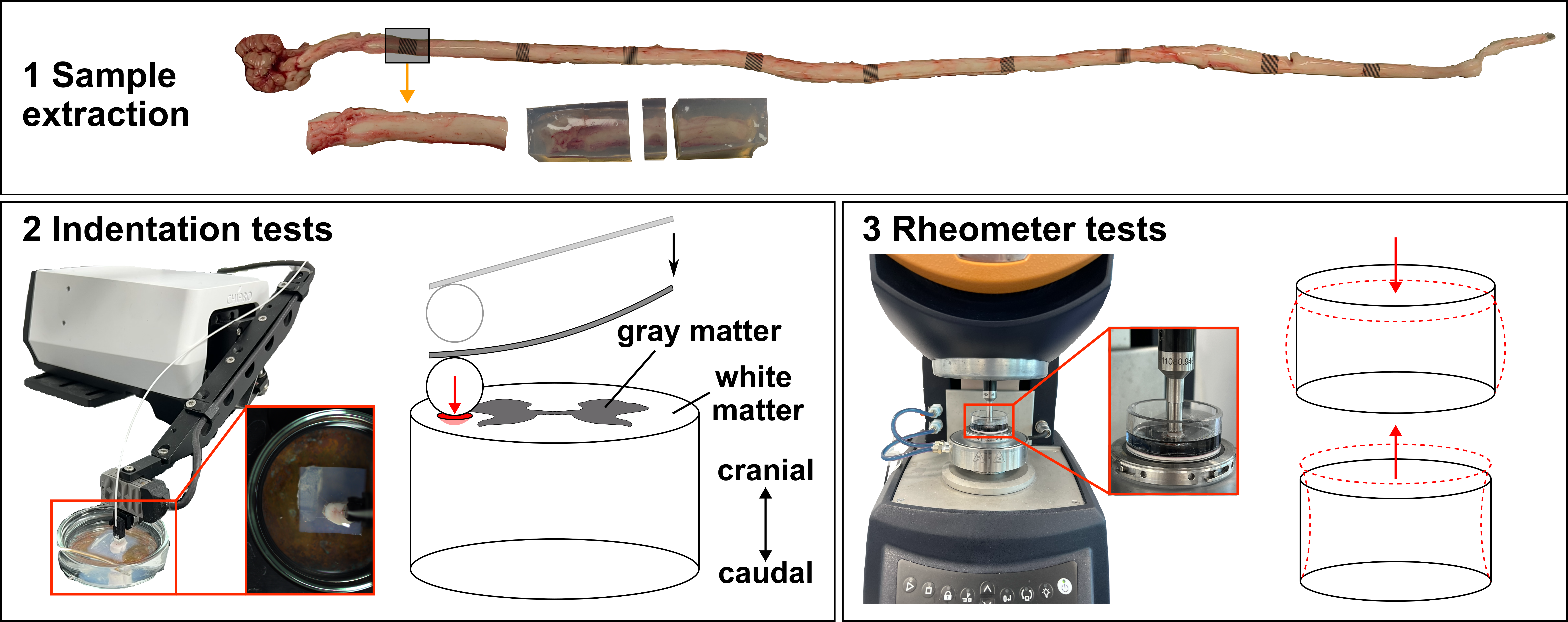
Figure 3. Multimodal experimental pipeline for spinal cord tissue characterization. (1) A segment was extracted from the porcine spinal cord, embedded, and dissected into individual samples. (2) Spherical indentation tests were performed on gray and white matter in the transverse plane using a Chiaro Nanoindenter. (3) The same samples were subsequently tested using a rheometer under large-strain compression and tension.
Using nonlinear continuum mechanics models to analyze both the indentation and rheometer force-displacement data, we identified complementary mechanical parameters for spinal cord gray and white matter [3]. This framework bridges mesoscale, tissue-resolving indentation with macroscale bulk mechanical testing and enables the identification of more robust, multimodally informed material parameters for computational studies of spinal cord biomechanics.
References:
- (1) Neumann, O.; Surana, H. V.; Melly, S. K.; Steinmann, P.; Budday, S. Mechanical Characteristics of Spinal Cord Tissue by Indentation. J Mech Behav Biomed Mater 2025, 163, 106863. https://doi.org/10.1016/j.jmbbm.2024.106863.
- (2) Neumann, O.; Surana, H. V.; Hintze, M.; Kuerten, S.; Franz, T.; Gopalan Ramachandran, R.; Steinmann, P.; Budday, S. Spatial Scale of Indentation Explains Shift in Ratio between Spinal Cord Gray and White Matter Stiffness. Sci Rep 2026, 16, 16989. https://doi.org/10.1038/s41598-026-55065-z.
- (3) Neumann, O.; Gopalan Ramachandran, R.; Surana, H. V.; Paulsen, F.; Scholz, M.; Gaffling, S.; Steinmann, P.; Budday, S. Multimodal Mechanical Characterization Pipeline for Spinal Cord Tissue. Acta Biomater 2026, 216, 248–271. https://doi.org/10.1016/j.actbio.2026.04.036.
About the authors

Oskar Neumann
Oskar Neumann is a doctoral researcher at the Institute of Continuum Mechanics and Biomechanics at Friedrich-Alexander-Universität Erlangen-Nürnberg (FAU), Germany, and a member of the Collaborative Research Centre 1540 “Exploring Brain Mechanics.”
With a background in engineering and mechanics, his research combines experimental tissue characterization with computational modeling to investigate the biomechanics of the healthy and regenerating spinal cord. His experimental work focuses on understanding how tissue microstructure, testing conditions, and spatial scale influence the mechanical properties of spinal cord gray and white matter.
Interested in presenting your research in Faces of Mechanobiology?
Send an email to marketing@optics11life.com
Mentioned in this article:
Chiaro
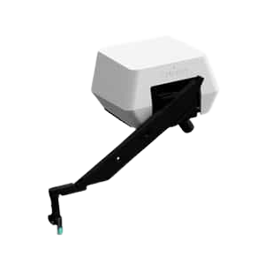
Chiaro is a microscope-compatible, nanoindentation system that allows you to combine unique mechanical insights with the imaging equipment of your choice. A compact yet powerful instrument to expand your lab infrastructure.