In this episode of Speaking from the Heart, Prof. Michael Monaghan of Trinity College Dublin discusses how engineered heart tissues can help researchers understand cardiac biology, investigate biomaterial influences, and explore new approaches to heart tissue repair.
The conversation explores how human induced pluripotent stem cell (iPSC)-derived cardiomyocytes, advanced biomaterials, and 3D engineered heart tissues can be combined to study tissue maturation, metabolism, and contractile function. A central question is how researchers can move beyond conventional cell models to understand how cardiac tissue responds to mechanical cues, electrical stimulation, and potential treatments.
Michael also shares how his group uses Cuore to monitor engineered heart tissue over time, investigate force kinetics and drug responses, and explore what’s needed to make advanced human cardiac models more reproducible, informative, and relevant to future therapies.
Watch the conversation here:
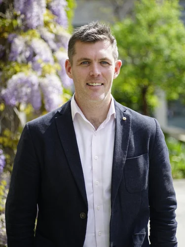
Prof. Michael Monaghan
Prof. Michael Monaghan is Professor of Biomedical Engineering at Trinity College Dublin, Ireland, in the Department of Mechanical, Manufacturing and Biomedical Engineering.
His research focuses on biomaterials, cardiac tissue regeneration, and advanced human tissue models, particularly those derived from induced pluripotent stem cells. His group combines biomaterials engineering, stem cell biology, and advanced imaging methods to investigate cardiac tissue development, cell metabolism, and interactions between cells and their mechanical environment.
His research also includes electroconductive biomaterials, fluorescence lifetime imaging microscopy (FLIM), and developing engineered heart tissues to study cardiac function.
By bringing together biomaterials research and functional human tissue models, Michael’s group aims to improve our understanding of cardiac biology while developing materials and approaches that could ultimately support heart repair.
What makes an engineered heart tissue more representative of the human heart?
Creating a 3D cardiac tissue model is an important step toward reproducing human heart biology, but cardiomyocytes alone do not necessarily capture the complexity of living heart tissue.
Cardiomyocytes interact with other cell types, respond to their surrounding extracellular matrix, and undergo changes in metabolism and structure as they mature.
For Michael’s group, this creates several important research questions. How does changing the cellular composition of an engineered heart tissue affect its maturation? What happens when different biomaterials are introduced? And how do these changes influence the tissue’s ability to contract?
The group uses human iPSC-derived cardiomyocytes to create engineered heart tissue and investigates how different supporting materials, cell populations, and culture conditions influence its development.
The goal is not simply to create a more complex model, but to understand which biological and mechanical factors contribute to a more functional human cardiac tissue.
Why does the move from 2D to 3D matter for cardiac biomaterials research?
Not every scientific question requires a 3D model.
As Michael explains, 2D cultures remain valuable for initial screening. They are generally less expensive, easier to work with, and suitable for evaluating the early effects of different materials.
But when researchers want to understand how biomaterials influence the mechanical behavior of cardiac tissue, the limitations of 2D systems become more apparent.
In particular, some materials have anisotropic mechanical properties, meaning their behavior depends on the direction of applied force.
3D engineered heart tissues provide a more suitable environment for investigating these properties. Cardiomyocytes can organize and align along the mechanical constraints created by the tissue supports, while researchers can examine how different materials influence tissue structure and contractile function.
The question, therefore, is not whether 3D should replace 2D, but when the added complexity of 3D provides information that simpler models cannot.
What can continuous contractility measurements reveal about cardiac tissue?
A cardiac tissue model can provide valuable information about cell composition, morphology, and molecular behavior. But understanding how that tissue functions requires researchers to measure its contraction.
And the amount of force generated is only part of the story.
Contraction dynamics, including time to peak force, relaxation, and changes in the contraction curve, can help researchers understand how cardiac tissues respond to their environment.
Michael’s group uses Cuore to culture multiple engineered heart tissues in parallel and continuously monitor their contractile behavior as they mature.
This is particularly relevant when studying how different biomaterials influence cardiac function or how established drugs affect contractile responses.
Rather than relying only on measurements at individual time points, continuous functional monitoring lets researchers observe how responses develop over time.
It also enables researchers to link changes in tissue mechanics with biological observations, providing a more complete understanding of how engineered cardiac tissues behave.
How do biomaterials and metabolism influence cardiac tissue maturation?
Cardiac maturation is not only a structural process. It also involves changes in how cardiomyocytes produce and use energy.
Michael’s group investigates these changes using fluorescence lifetime imaging microscopy, which lets researchers study cellular metabolism during cardiomyocyte differentiation and maturation.
At the same time, the group develops biomaterials that provide different stimuli to engineered heart tissues.
This raises an important question: how do changes in the mechanical and biochemical environment influence metabolism, maturation, and ultimately cardiac function?
The group has already observed differences in contractile force and drug responses between engineered heart tissues exposed to different metabolic maturation conditions.
Interestingly, some responses have not followed the patterns the researchers initially expected.
These observations highlight why advanced models are valuable for more than confirming existing assumptions. They can also reveal biological behavior that requires further investigation.
Can more standardized biomaterials improve the reproducibility of cardiac models?
As advanced human models become more widely used, reproducibility remains an important challenge.
Different laboratories work with different iPSC lines, differentiation protocols, and culture conditions. Even when researchers follow similar procedures, the resulting engineered tissues may not behave the same way.
The extracellular matrix is another important variable.
Because the matrix provides the physical environment surrounding the cells, differences in its composition and mechanical properties can influence tissue behavior.
Michael suggests that developing synthetic, standardized extracellular matrices could help reduce some of this variability and make results more reproducible across laboratories.
However, standardizing the matrix alone will not solve every challenge. Differences in cell lines, differentiation success, and experimental procedures must also be considered.
For engineered heart tissues to become more widely adopted in research and drug development, improving reproducibility will be just as important as increasing biological complexity.
How much complexity do engineered heart tissues need?
One direction in advanced cardiac model development is adding more cell types.
The heart contains much more than cardiomyocytes. Immune cells, for example, can influence inflammation, tissue remodeling, and injury responses.
Michael explains how his group is already incorporating innate immune cells, including macrophages, into engineered heart tissue models. Looking ahead, researchers may increasingly incorporate other immune populations, including cells involved in adaptive immune responses.
Another challenge arises as models grow larger.
As engineered tissues grow, the need for vascularization becomes increasingly important. Without an adequate supply of oxygen and nutrients, cells in the center of larger tissues may become damaged or die.
These challenges raise a broader question for the field: how can researchers make models more representative of human tissue while keeping them practical, reproducible, and suitable for experimentation?
More complexity may improve biological relevance, but it also introduces new technical and experimental challenges.
What comes next for functional cardiac tissue monitoring?
Continuous contractility measurements already provide valuable insights into engineered heart tissue function.
But what if researchers could follow several aspects of tissue biology at the same time?
Michael sees an opportunity to expand functional monitoring beyond contraction force to include additional metabolic and biochemical measurements.
Parameters such as oxygen consumption, lactate production, glucose utilization, and fatty acid metabolism could provide complementary information about how cardiac tissues develop and respond to different conditions.
This becomes particularly interesting when studying cardiac injury.
Rather than examining a tissue only at the end of an experiment, researchers could potentially monitor how an injury develops, how tissue function changes, and how those changes respond to different treatments.
The ambition is to move toward more multiparametric models that provide a continuous picture of cardiac tissue behavior, combining mechanical function with additional biological information.
From engineered heart tissues to cardiac repair
Beyond developing better in vitro models, Michael’s group has a longer-term interest in translating biomaterials research toward cardiac repair.
Following a myocardial infarction, damaged heart tissue undergoes substantial changes, including inflammation, remodeling, and scar formation.
New materials could potentially influence these processes by modifying the local environment, reducing inflammation, or promoting wound healing.
Michael describes two complementary directions in his research.
The first is developing more representative human cardiac models that allow researchers to investigate tissue function and evaluate different materials and stimuli.
The second is developing acellular biomaterials that could eventually be applied to damaged heart tissue in a clinical setting.
Engineered heart tissues provide a way to investigate these approaches in vitro before considering their potential therapeutic applications.
The longer-term goal is to connect a better understanding of cardiac biology with the development of materials that may one day support recovery after heart injury.
Would you like to participate in Speaking from the Heart?
Email us at marketing@optics11life.com and tell us about your research.