by Elizaveta Loseva and Giulia Pilia (Optics11 Life)
© 2026 Optics11 Life B.V
Muscle contractility is a fundamental functional parameter in muscle physiopathology. Traditionally, contractility has been studied in vivo, posing questions about intra-species translatability and raising ethical and cost-related concerns, or in 2D cell culture, with limited physiological relevance. In recent years, the development of methods for the generation of engineered muscle tissue has opened the way to model muscle function in vitro employing human cells, retaining the 3D architecture and allowing for quantitative analysis of force generation. However, such assays are often complex to develop and labor-intensive, impairing their scalability and reproducibility.
In this application note, we present an assay for the functional evaluation of an iPSC-derived skeletal muscle model in Cuore, a platform enabling generation and contractility testing of 3D muscle tissues. Showcasing the long-term monitoring of the model evolution over time, its response to drug treatment, and downstream analysis of the 3D constructs, we provide the blueprint for the design and customization of diverse functional assays.
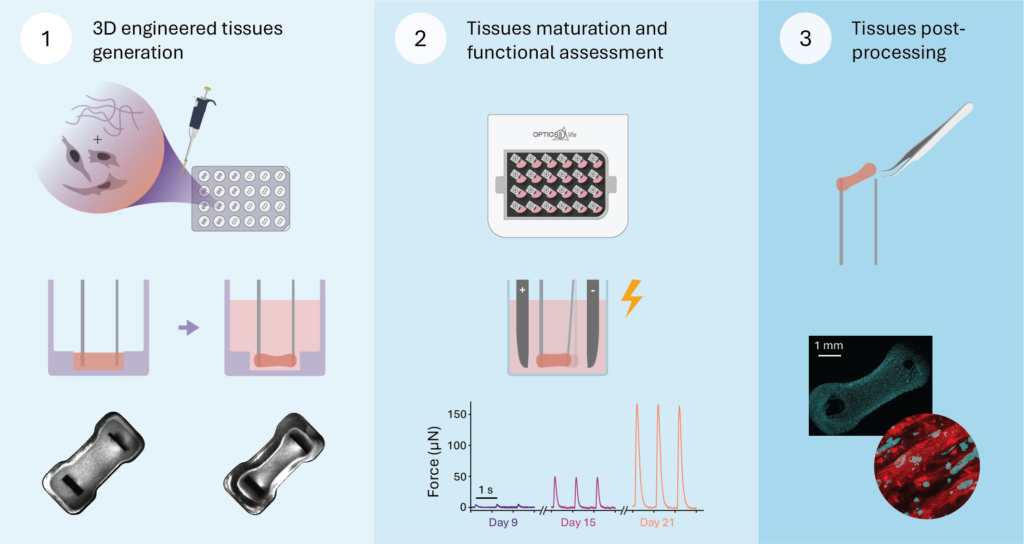
Figure 1
Overview of the presented experimental workflow. (1) Muscle tissues are generated by mixing cells with hydrogel and dispensing them into a 24-well casting mold, followed by tissue compaction around a pair of cantilevers. (2) The casting mold is replaced by a well plate, and a stimulation plate is added into the assembled smart lid for electrical pacing of the tissues. Graphs at the bottom demonstrate twitch force increase over time for one example tissue. (3) At the end of the experiment, tissues can be harvested and used for downstream analyses. Microscopy image shows a whole tissue stained with Hoechst; zoom-in view: multinucleated striated fibers (cyan – nuclei, red – titin)
Results and Methods
Workflow overview
3D engineered skeletal muscle tissues were generated as described in Iuliano et al.,1 employing hiPSC-derived myogenic progenitors kindly provided by prof. Pim Pijnappel and derived as described in van der Wal et al.2, 3 In brief, 6·105 cells per tissue were resuspended in standard growth medium and incorporated in a hydrogel mix composed of 2 mg/ml fibrinogen, 0.5 U/ml thrombin and 20% growth factor reduced Matrigel, to a total volume of 50 µl. Following the dispensing of the cell-hydrogel mixture into the Cuore’s casting mold and assembling the smart lid, tissue compaction around the cantilevers was monitored (Figure 1, left panel). For the first 2 days, tissues were grown in standard growth medium containing 1% antibiotic-antimycotic solution and 1.5 mg/ml 6-aminocaproic acid. Two days upon casting, the tissues achieved sufficient compaction and were transferred from the casting mold into a standard 24-well plate containing differentiation medium (DMEM 1 g/L glucose, 1% Knock-Out Serum Replacement, 1% antibiotic-antimycotic solution, 2 mg/ml 6-aminocaproic acid). This moment marks the initiation of the final differentiation process and is referred to as “day 0”. The medium was refreshed on day 3 and every second day thereafter.
In the following days, the cells fused and matured into multinucleated myofibers, acquiring the ability to contract as a coherent tissue. Starting from day 7 and for successive 14 days, functional evolution of the tissues was followed by non-destructive monitoring of contraction forces and kinetics every second day (Figure 1, middle panel; Figure 2). Both low-frequency (1 Hz) and high-frequency (50 Hz) stimulation were applied to induce twitch and fused tetanic contractions, respectively. At the experimental endpoint, the architecture of our 3D constructs was assayed by immunostaining the structural protein titin and imaging the whole tissue (Figure 1, right panel). The presence of numerous long multinucleated myofibers, and the characteristic striated structure of the sarcomere could be observed.
Detailed insight on tissue contractility
For each measurement day, the force-time tracks obtained upon twitch (10 s of 1 Hz stimulation at 3.5V, with a pulse width of 5 ms in bipolar mode) and tetanic stimulation (three 1 s-long trains of 50 Hz stimulation at 3.5 V, with a pulse width of 5 ms in bipolar mode and 30 s spacing between the trains) were analyzed. The absolute expressed force, as well as several contraction kinetic parameters, were extracted (Figure 2). This detailed analysis is a default in Prova, the analysis software integrated in the Cuore package.
A stable maximum contraction force recorded for the first few measurements grew progressively in the following days (Figure 2a). This trend was consistent for both twitch and tetanic contractions, with tetanic contractions being, as expected, considerably stronger. The velocity of contraction and relaxation for both twitch and tetanic contractions increased progressively over time (Figure 2b, c). However, while the evolution of relaxation velocity during twitch contraction was still showing an upward trend at the experimental endpoint, that of tetanic contractions reached a plateau from day 17.
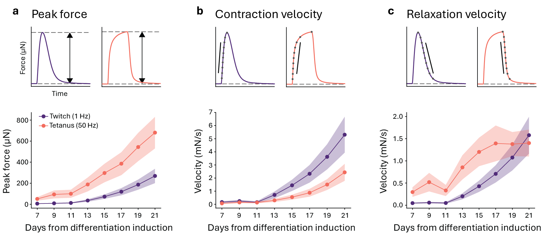
Figure 2
Evolution of twitch and tetanic contraction parameters over the course of the experiment. (a) Peak contraction force. (b) Contraction velocity measured as linear slope of the curve between 20% and 80% of peak force. (c) Relaxation velocity measured as linear slope of the curve between 80% and 20% of peak force. Solid line: mean value; shading: standard deviation (n = 21 tissues).
Response to drug administration
The assessment of functional drug effects in vitro is fundamental to reduce, refine, and replace animal testing. To validate the use of Cuore in this context, two reference compounds with known effects on muscle contractility, caffeine and verapamil, were tested. Caffeine was dissolved in ultrapure water at 50 mM and administered at final concentration of 0.1 mM or 1 mM. Verapamil was dissolved in DMSO at 10 mM and administered at 1 μM or 10 μM. Drug administration was performed on day 21 of the experiment, by swapping the well plate for one containing pre-warmed differentiation medium with either drug or vehicle control. Wash-out was performed by immersing the tissues in warm DMEM (1 g/l glucose) for 1 minute and then placing them in a well plate with warm differentiation medium. Baseline contractions were measured for 5 minutes before the plate swap, and then the onset of acute functional effect upon drug administration and following wash-out were monitored, by continuously stimulating the tissues with five 1 Hz pulses at 3.5 V once a minute.
As expected, caffeine induced rapid increase of contraction forces, which was dose-dependent and sustained in time (Figure 3a). The transient effect of caffeine was promptly reverted by the wash-out, and previously treated tissues recovered normal function. Mechanistically, caffeine increases muscle contractility by increasing the sensitivity of the ryanodine receptor and therefore enhancing calcium release from the sarcoplasmic reticulum4. Reflecting the molecular mechanism of action, caffeine-treated tissues contracted faster than the untreated controls (Figure 3b), while the effect on the relaxation velocity was milder (Figure 3c).
While primarily used to treat cardiovascular conditions in the clinic, verapamil also attenuates skeletal muscle contractility at higher concentrations, by impairing sarcoplasmic calcium release through both direct and indirect mechanisms.5, 6 In our experiments, a stark dose-dependent inhibition of contraction force was observed, with rapid onset upon drug administration (Figure 3d). This effect was not reversed by drug removal in the time considered, indicating slow dissociation kinetics. Verapamil’s treatment induced slower contraction rates (Figure 3e), coherently with its mechanism of action. Additionally, the velocity of tissue relaxation was also reduced (Figure 3f), in line with previous findings that verapamil can also impair calcium re-uptake7.
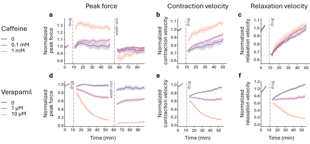
Figure 3
Changes in twitch peak force, contraction velocity (between 20% and 80% of maximum force) and relaxation velocity (between 80% and 20% of maximum force) upon caffeine (top panels) and verapamil (bottom panels) administration. Force and velocity values for each tissue are normalized to the respective value before drug administration. Dotted vertical lines indicate the time of substituting the well plates. Solid line: mean value; shading: standard deviation (n = 21 tissues in caffeine experiment (n=8, 0 mM; n=7, 0.1 mM; n=6, 1 mM); 20 tissues in verapamil experiment (n=6, 0 μM; n=6, 1 μM; n=8, 10 μM)).
Conclusions
- Cuore enables integrated workflows for the study of skeletal muscle function in vitro, as exemplified by the presented setup employing hiPSC-derived 3D engineered tissues.
- Our platform allows monitoring the evolution of tissue contractility longitudinally for extended windows of time. We terminated our experiment after two weeks of functional measurements, when tissues were still intact and responsive, indicating that longer experimental design is possible.
- The presented workflow is compatible with testing acute drug effects. Using reference compounds with known effects on muscle contraction, we captured onset of drug response and its reversibility, as well as changes in contraction dynamics suggestive of the mechanism of action of the drug.
- Long-term contractility monitoring, together with the possibility of downstream analyses of the tissue (e.g. imaging, quantification of gene expression or protein content) enables a thorough characterization of the model. Facilitating the assessment of the chronic effects of genetic manipulation or compound treatments, this integrated approach has broad applicability in disease modeling and toxicology.
References
- A. Iuliano et al. Real-time and Multichannel Measurement of Contractility of hiPSC-Derived 3D Skeletal Muscle using Fiber Optics-Based Sensing. Adv. Mater. Technol. 8, 22, 2300845 (2023).
- E. van der Wal et al. Large-Scale Expansion of Human iPSC-Derived Skeletal Muscle Cells for Disease Modeling and Cell-Based Therapeutic Strategies. Stem Cell Rep. 10, 6, 1975–1990 (2018).
- E. van der Wal et al. Highly contractile 3D tissue engineered skeletal muscles from human iPSCs reveal similarities with primary myoblast-derived tissues. Stem Cell Rep. 18, 10, 1954–1971 (2023)
- T. Murayama, H. Ogawa, N. Kurebayashi, S. Ohno, M. Horie & T. Sakurai. A tryptophan residue in the caffeine-binding site of the ryanodine receptor regulates Ca2+ sensitivity. Commun. Biol. 1, 1, 98 (2018)
- H. H. Valdivia, C. Valdivia, J. Ma & R. Coronado. Direct binding of verapamil to the ryanodine receptor channel of sarcoplasmic reticulum. Biophys. J. 58, 2, 471–481 (1990).
- F. S. Menezes-Rodrigues, M. Pires-Oliveira, T. Duarte, E. J. Paredes-Gamero, T. Chiavegatti & R. O. Godinho. Calcium influx through L-type channels attenuates skeletal muscle contraction via inhibition of adenylyl cyclases. Eur. J. Pharmacol. 720, 1–3, 326–334 (2013).
- M. J. Paydar, A. Pousti, H. Farsam, M. Amanlou, S. E. Mehr & A. R. Dehpour. Effects of diltiazem or verapamil on calcium uptake and release from chicken skeletal muscle sarcoplasmic reticulum. Can. J. Physiol. Pharmacol. 83, 11, 967–975 (2005)