Native tissues do not behave as purely elastic materials. Their mechanical response changes over time, meaning that cells encounter not only stiffness, but also stress relaxation and energy dissipation in their surrounding microenvironment.
In this Faces of Mechanobiology entry, Mariana A.G. Oliva and Manuel Salmeron-Sanchez explore why viscoelasticity is an important consideration when building more physiologically relevant in vitro models. They discuss how a tunable hydrogel platform was used to independently control stiffness and stress relaxation, how microscale nanoindentation characterisation supported the comparison of these materials, and how computational modelling helped reveal the role of Piezo1 in mechanotransduction within soft, dissipative matrices.
Understanding cell response to dynamic tissues
Tissues in the body don’t behave like elastic solids, and their mechanical properties change over time when deformed. Indeed, it is increasingly understood that tissues in the body are dynamic and that their response to applied deformation is a time-dependent process. Advances in the fields of biomaterials have allowed us to replicate these previously unexplored properties in-vitro, providing advanced materials that can both replicate specific tissue stiffness and dynamic properties such as stress relaxation (viscoelasticity). Still, there is much to explore regarding how cells perceive dynamic properties and how these guide cell behaviour. A promising new study1 investigated how mesenchymal stem cells (MSCs) sense time-dependent cues, revealing Piezo1 as a key player in soft matrix mechanotransduction.
Key aspects:
Defining Viscoelasticity
A key aspect of this work was to recapitulate native tissue viscoelasticity in a tuneable, yet minimalistic, hydrogel platform. While elasticity (defined by the elastic modulus) is a standardized metric, often measured via AFM, nanoindentation or rheology; viscoelastic properties lack such a consensus, as they vary with timescale, frequency, and method. A hydrogel might seem rigid under fast nanoindentation but fluid over hours, muddling cross-study comparisons. This is a key challenge in mechanobiology: without shared definitions, it is difficult to link matrix properties to cell behaviour. In this study, hydrogel characterisation was mainly performed by nanoindentation, using the Chiaro nanoindenter2. The instrument allowed for the microscale characterisation of all hydrogels employed, including elastic modulus, stress relaxation (viscoelasticity) behaviour, as well as topography, all important metrics when considering material characterisation and controlled cell-material interactions. From these metrics, it was possible to discern hydrogel groups with independently tuneable elastic and viscoelastic properties: soft and stiff hydrogels groups with no significant difference in terms of Young’s modulus ( E ~ 0.4 and 25 kPa, respectively) but with an elastic (V-, slow stress relaxing) and viscoelastic (V+, fast stress relaxing) counterpart.
Matching in vitro data with computational modelling
Another key consideration was understanding how cells respond to ‘soft’ and ‘stiff’ viscoelasticity. Researchers approached this question by performing experiments with an immortalized MSC line and a modified computational clutch model, which aims to predict how integrins will bind (clutch engaged) the substrate they are on. Using the hydrogel mechanical measurements (E and relaxation time constant, 𝜏) as inputs for model parameters, the team expanded existing models3 to explore how varying viscoelasticity could influence cell-material interactions. Additionally, researchers added the activity of the mechanosensitive ion channel Piezo1 (identified as an integrin activator4) to the computational model. The model encompassed for the first time the interaction between Piezo1, integrins, and viscoelastic substrates, and it confirmed experimental findings, which proposed Piezo1 as a crucial regulator of cell mechanotransduction in soft dissipative matrices (Figure 1).
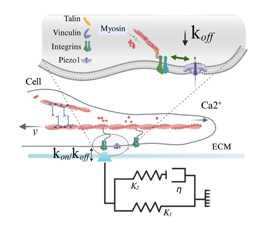
Figure 1. Schematic of the influence of Piezo1 in molecular clutch engagement. Cell is shown to be coupled to the ECM via ECM-binding integrins that in turn, connect to the contractile actin filaments via mechanosensitive adaptive proteins (talin and vinculin). Myosin motors continuously pull on actin filaments with velocity (v). Here, the ECM is modelled as a Standard Linear Solid (SLS), composed of two elastic springs (K1 and K2) and a viscous dashpot element (η). kon and koff represent the rates of clutch association and dissociation, respectively. In the zoom (grey shading), the concerted action between Piezo1 and integrins is highlighted, showing that potentiation of this interaction decreases clutch dissociation (koff), adapted from 1.
Take home message
The dynamic, time-dependent properties of tissues are thus an important metric to consider when building relevant physiological in-vitro models. The work reported here highlights not only the importance of matrix viscoelasticity in modulating cell behaviour but also how cell response to substrate energy dissipation is dependent on the stiffness range, with enhanced viscoelasticity potentiating cellular ‘mechanoactivation’ in soft regimes, and the opposite above a stiffness threshold (see 5). Soft tissues in the body, such as brain, lung tissue, or bone marrow, exhibit the highest degree of energy dissipation, and many of the existing in-vitro models that aim to replicate these environments are designed with only elastic properties in mind. Finally, authors highlight how the Nobel Prize-recognized mechanosensation pathway involving Piezo1 provides a potential mechanistic framework for Piezo1’s involvement in neural tissue physiology and pathophysiology.
References:
- A. G. Oliva, M. et al. Piezo1 regulates the mechanotransduction of soft matrix viscoelasticity. Nat Commun 16, 9155 (2025).
- Ciccone, G. et al. Experimental and Data Analysis Workflow for Soft Matter Nanoindentation. JoVE (Journal of Visualized Experiments) e63401 (2022) doi:10.3791/63401.
- Elosegui-Artola, A. et al. Mechanical regulation of a molecular clutch defines force transmission and transduction in response to matrix rigidity. Nature Cell Biology 18, 540–548 (2016).
- McHugh, B. J. et al. Integrin activation by Fam38A uses a novel mechanism of R-Ras targeting to the endoplasmic reticulum. Journal of Cell Science 123, 51–61 (2010).
- Chaudhuri, O. et al. Substrate stress relaxation regulates cell spreading. Nature Communications 6, 6365 (2015).
About the authors

Mariana Azevedo Gonzalez Oliva
Mariana Azevedo Gonzalez Oliva is a researcher in the Microenvironments for Medicine group at the Institute for Bioengineering of Catalonia, IBEC. Her work focuses on how cells sense and respond to the mechanical properties of their environment, including matrix stiffness, viscoelasticity, and stress relaxation.

Manuel Salmeron Sanchez
Manuel Salmeron-Sanchez is an ICREA Research Professor and Group Leader of the Microenvironments for Medicine group at IBEC. His research focuses on advanced biomaterials for cell and tissue engineering, with the goal of engineering biochemical and biophysical cues in the cellular microenvironment for mechanobiology and regenerative medicine applications.
Learn more about the Institute for Bioengineering of Catalonia (IBEC) here.
Interested in presenting your research in Faces of Mechanobiology?
Send an email to marketing@optics11life.com
Mentioned in this article:
Chiaro
Chiaro is a microscope-compatible, nanoindentation system that allows you to combine unique mechanical insights with the imaging equipment of your choice. A compact yet powerful instrument to expand your lab infrastructure.